Urology

Department Page

About Urology

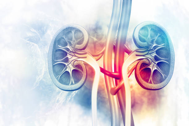
The urinary tract is your body’s drainage system for removing urine. Urine is composed of wastes and water. The urinary tract includes your kidneys, ureters, and bladder. To urinate normally, the urinary tract needs to work together in the correct order.

Urologic diseases or conditions include urinary tract infections, kidney stones, bladder control problems, and prostate problems, among others. Some urologic conditions last only a short time, while others are long-lasting.

Indira Super Speciality Hospital

SYMPTOMS :

  1. Burning Sensation when passing urine
  2. Frequent urination
  3. Pain or pressure in the lower Abdomen
  4. Dark / Cloudy urine
  5. Fever / Chills

Risk Factors :

  1. Obstruction in the urinary Tract- Stones / Enlarged prostate
  2. Diabetes
  3. Postmenopausal women due to hormonal changes
  4. Elderly men due to poor Bladder emptying
  5. Immunocompromised State

Diagnosis :

  1. Urine Analysis
  2. Urine Culture
  3. Imaging of urinary Tract – Ultrasound
  4. Cystoscopy in case of Recurrent UTI
  5. Rule out Diabetes

Diagnosis :

  1. Antibiotics
  2. Analgesics
  3. Vaginal estrogens in postmenopausal women

Symptoms of Urinary Incontinence :

  1. Sui – Leakage of urine due to increase in Abdominal Pressure (E.G.) during Coughing, Sneezing , Laughing, Exercising or Lifting Weight.

Urge Incontinence :

  1. Sudden intense urge to urinate followed by an involuntary loss of urine

Overflow incontinence :

  1. Frequent or constant dribbling of urine due to a bladder that does not empty completely

Mixed Incontinence :

  1. More than one type of urinary incontinence
  2. Temporary Incontinence can be caused by intake of alcohol, Caffeine, Carbonated Drinks, Sweetness, Citrus Fruits, Heart and Blood Pressure Medications etc.
  3. Other causes are UTI, constipation, pregnancy, childbirth, menopause, hysterectomy enlarged prostate, prostate cancer, obstruction, Neurological Disorders, Multiple Sclerosis, Parkinson’s Disease, Stroke, Spinal Injury etc.

Treatment :

Depends on the type of incontinence:

  1. Behavioral Techniques
    1. Bladder Training
    2. Double Voiding
    3. Scheduled toilet trips
    4. Fluid and Diet management
  2. Pelvic floor exercises
  3. Electrical Stimulation
  4. Medications EG. Anticholinergics
  5. Medical Devices
    1. Urethral Insert
    2. Pessary
    3. Scheduled toilet trips
    4. Fluid and diet management
  6. Interventional Therapies
    1. Bulking material injection
    2. Botulinum Toxin(BOTOX)
    3. Nerve Stimulators
  7. Surgery
    1. Sling procedures
    2. Bladder neck suspension
    3. Prolapse Surgery
    4. Artificial urinary sphincter
  8. Absorbent Pads / Catheters

Phimosis :

It is the inability to retract the foreskin back from the head of the penis.

Causes :

  1. Scar Tissue
  2. Medical conditions like Diabetes / Balanitis

Symptoms :

  1. Difficulty in passing urine / burning sensation
  2. Discharge

Diagnosis :

  1. Clinical Diagnosis

Treatment :

  1. Circumcision

It is the narrowing of the urethra caused by injury, Instrumentation, Infection and certain non infectious forms of Urethritis.

Causes :

  1. Trauma
  2. Instrumentation
  3. Infection
  4. Post surgery done for hypospadias

Complications :

  1. Retention of urine
  2. Urinary Infection
  3. Bladder Dysfunction
  4. Bilateral Hydronephrosis

Diagnosis:

  1. Cystoscopy
  2. Ascending Urethrogram

Treatment :

  1. Dilation and other Endoscopic approaches
  2. Urethroplasty
  3. Urethral Stent

It involves hyperplasia of prostatic cells, resulting in the formation of large, fairly discrete nodules in the transition zone of the prostate.

Signs and Symptoms :

  1. Frequent Urination
  2. Urgency
  3. Involuntary Urination
  4. Urge Incontinence
  5. Pain on passing urine

Differential Diagnosis :

  1. Urinary Tract Infection
  2. Urethral Stricture
  3. Urethral Calculi
  4. Chronic Prostatis
  5. Bladder Cancer
  6. Prostate Cancer

DIAGNOSIS :

  1. Per Rectal Examination – Enlarged Prostate
  2. Urinalysis
  3. Prostate Specific Antigen (PSA)
  4. Transrectal / Tranabdominal Ultrasonography
  1. Medications :
    1. Alpha Adrenergic Agonist
    2. Anticholinergic Medications
  2. Self Catheterization
  3. Surgery
    1. Turp – Transurethral resection of the Prostate
    2. Open Prostatectomy – not usually performed now a days
    3. Holmium Laser Enucleation of the Prostate (HOLEP)

It is an abnormal enlargement of the venous plexus, in the scrotum. Occurs in 15%.

Signs and Symptoms :

  1. Soft lumps above the testicles
  2. Pain / Heaviness in the Scrotum
  3. Male Infertility

Diagnosis :

  1. By ultrasound showing dilation of the vessels greater than 2 mm. THE VESSELS GREATER THAN 2 MM.

Treatment:

Surgical Approaches

  1. Retroperitoneal – Laparoscopic Surgery
  2. Inguinal – Using percutaneous embolization

It is stone lying within the ureter at any point from the upper to lower ureter.

Signs and Symptoms :

  1. Severe colicky pain in the loin
  2. Radiation of pain to the Groin, Perineum and Testis
  3. Vomiting
  4. Bloody Urine
  5. Fever with rigors (If there is infection)

Diagnosis :

  1. X-Ray KUB
  2. Ultrasonography
  3. CT Abdomen

Differential Diagnosis:

  1. Appendicitis
  2. Ovarian causes in women
  3. Salphingitis in women

Management :

  1. Pain Killers
  2. Plenty of Fluids
  3. Diuretics

Indications for Surgery :

  1. Persistent Loin pain
  2. Vomiting due to stone obstruction
  3. Failure of stone progression

Treatment

  1. Laser Lithotripsy

Symptoms :

  1. Lower Abdominal pain
  2. Burning while passing urine
  3. Interrupted urine flow
  4. Blood in urine / Cloudy Urine

Causes of Bladder Stones :

  1. Prostate gland enlargement can abstruct the flow of urine, preventing complete emptying of the Bladder.
  2. Neurogenic Bladder – Preventing Bladder Emptying.
  3. Medical Devices – Bladder catheters causes formation of mineral crystals which later become stones.

Risk factors for Bladder Stones :

  1. Bladder outlet obstruction
  2. Neurogenic Bladder

Complications :

  1. Chronic Bladder Dysfunction
  2. Urinary Tract Infection
  3. Carcinoma Bladder in long standing Erosion due to Bladder stone.

Diagnosis :

  1. X RAY – Some type of stones may not be visible on conventional X Rays
  2. Ultrasound
  3. CT – most sensitive test to identify small stones.

Treatment :

  1. Lithotripters / Laser Lithotripsy
  2. Surgical removal by making an incision in the Bladder.

Renal Stones are formed in the kidneys. They most often occur in the age group of 20 – 40 yrs.

Types of Renal Calculi :

  1. Calcium Stones – Oxalate, Phosphate eg. Those who take diet rich in Oxalates.
  2. Uric Acid stones eg. Those with gout.
  3. Struvite – due to infection most commonly UTI. eg. Potato chips, Nuts, Spinach.
  4. Cystine stones

Risk Factors :

  1. Family history
  2. Urinary Tract Abnormalities
  3. Diet – high levels of protein, salt and sugar
  4. Dehydration

Symptoms :

  1. Severe colicky pain
  2. Vomiting
  3. Immobilization
  4. Fever with Rigors

Diagnosis :

  1. Ultrasound
  2. IVP
  3. CT Scan

Treatment :

  1. Lithotripsy – ESWL
  2. Percutaneous Nephrolithotomy – PCNL
  3. Rirs

Prevention :

  1. To drink atleast 2.5 L of fluid / day.

Renal Tumor :

  1. They can be either benign or malignant tumors.
  2. Benign Tumors – Less Common.
  3. Eg. Renal Oncocytoma, Cystic Nephroma, Renal Angiomyolipoma, Medullary Fibroma.

Malignant Tumor :

  1. Renal Cell Carcinoma accounts for 90 – 95% of malignant neoplasms arising from the kidney

Risk Factors :

  1. Tobacco Chewing
  2. Obesity
  3. Occupational Exposure
  4. Polycystic Kidney
  5. Genetic

Signs and Symptoms :

  1. Hypertension
  2. Hypercalcemia
  3. Polycythemia mostly diagnosed insidentally

Classic Triad :

  1. Haematuria
  2. Abdominal pain
  3. Palpable mass is seen in ≤ 10 % of cases

DIAGNOSIS :

  1. Imaging Technology
  2. Ultrasound / CT with contrast
  3. Chest XRAY
  4. MRI to evaluate tumor extension into IVC
  5. Needle Biopsy if mass < 3 CMS.

Treatment :

  1. Radical Nephrectomy is the treatment of choice and depends on the stage of disease.
  2. Immunotherapy – Reserved for patients with a good prognosis.
  3. Chemotherapy – for treating metastatic diseases.

Signs and Symptoms :

  1. Excessive growth of facial & body hair
  2. Enlargement of penis in boys or clitoris in girls
  3. Puberty earlier than usual
  4. Enlargement of breast in male
  5. Weight gain
  6. Purple stretch marks on the Abdomen.
  7. Menstrual irregularities
  8. Depression
  9. High BP
  10. High Sugar

DIAGNOSIS :

  1. Ultrasound
  2. CT Scan
  3. MRI
  4. Image guided FNAC of the mass

Treatment :

  1. Adrenalectomy
  2. Radiation Therapy
  3. Chemotherapy.

Bladder cancer is the abnormal growth of bladder cells and is a common cancer.

Signs and Symptoms :

  1. Using Tobacco, especially smoking cigarettes.
  2. Having a family history of bladder cancer.
  3. Post Chemoradiation
  4. Drinking water that has been treated with chlorine
  5. Using urinary catheters for a long time.
  6. Older age is a risk factor for most cancers. The chance of getting cancer increases as you get older
  7. More common in men than women

Prevention :

  1. Avoid smoking
  2. Limit exposure to chemicals
  3. Intake of plenty of fluids
  4. Intake of plenty of fruits and vegetables

Signs and Symptoms :

  1. Passing blood in urine
  2. Frequency
  3. Pain and burning during urination
  4. Poor urine stream
  5. Difficulty in passing urine
  6. Loss of weight

Tests for Bladder Cancer :

  1. Ultrasound
  2. Contrast CT Abdomen
  3. Biopsy( Histo pathology from Bladder tissue)

Types of Bladder Cancer :

There are three types of Bladder cancer that begin in cells in the lining of the Bladder.

These cancers are named for the type of cells that become malignant (Cancerous) :

  1. Transitional Cell Carcinoma
  2. Squamous Cell Carcinoma
  3. Adenocarcinoma

Treatment :

  1. Superficial Bladder Tumor
    1. Transurethral resection of Bladder Tumor or “TURBT”.
  2. Invasive Bladder Tumor
    1. Ideal conduit
    2. Orthotopic Neobladder
    3. Continent Catheterizable pouch – A form of Neobladder
    4. Radical Cystectomy (Open, Laparoscopic, or Robot Assisted) Combined with one of the three urinary diversion methods is the gold standard
  3. Metastatic Tumor :
    1. Chemotherapy

It is a Malignant growth on the skin or in the tissues of the penis. 95% of penile cancers are squamous cell Carcinomas

Risk Factors :

  1. Infections
    1. HIV Infection – Eight fold increase
    2. HPV Infection – Contributes to 40% of Penile Cancers
    3. Genital Warts
  2. Hygiene and Injury :
    1. Poor Hygiene
    2. Smegma – A Whitish substance that can accumulate beneath the foreskin and increase the risk of Penile Cancer.
    3. Balanitis and Penile Injury – Inflammation of the foreskin and the glans Penis, increases risk. Allergic reactions to soaps, infection or diabetes can predispose.
  3. Other Causes – Age more than 55 yrs; Lichen sclerosus; Tobacco smoking, ultraviolet light increases risk of penile cancer.

Prevention :

  1. HPV Vaccine
  2. Use of condoms – Acting protective against HPV.
  3. Good Genital hygiene.
  4. Cessation of smoking
  5. Circumcision during infancy or childhood.
  6. Preventing phimosis and paraphimosis

Treatment :

  1. Surgery: Amputation of the penis – Partial or total.
  2. Radiation Therapy
  3. Chemotherapy

Prognosis :

Earlier the cancer is diagnosed, the better the prognosis.